
Part 3 / 3
ADNEXAL ULTRASOUND: Benign and malignant adnexal tumors
The third course in a comprehensive three-part curriculum for Adnexal Ultrasound
from Physiology to Oncology
UZSСHOOL
Where Ultrasound Meets Surgery
Who Is This Course For?
Ultrasound Specialists
Obstetricians & Gynecologists
Radiologists
Reproductive Medicine Specialists
Gynecologic Oncologists
Residents & Sonographers
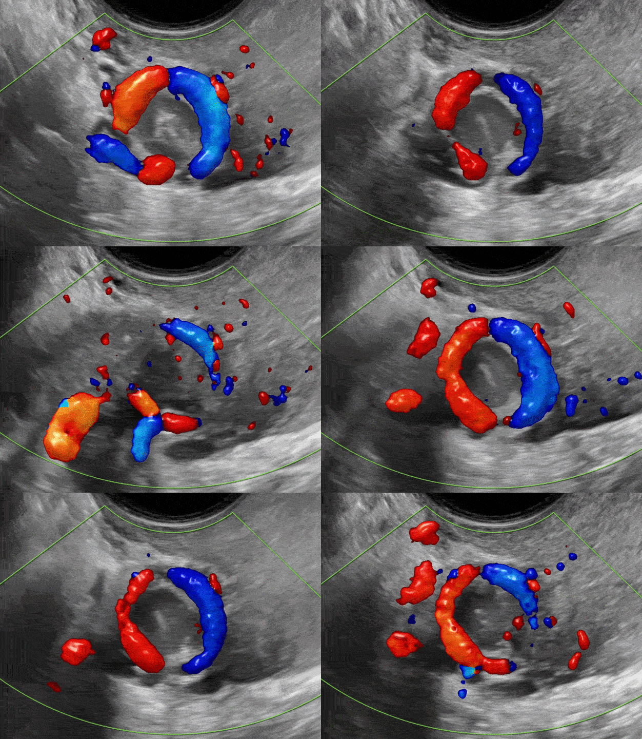
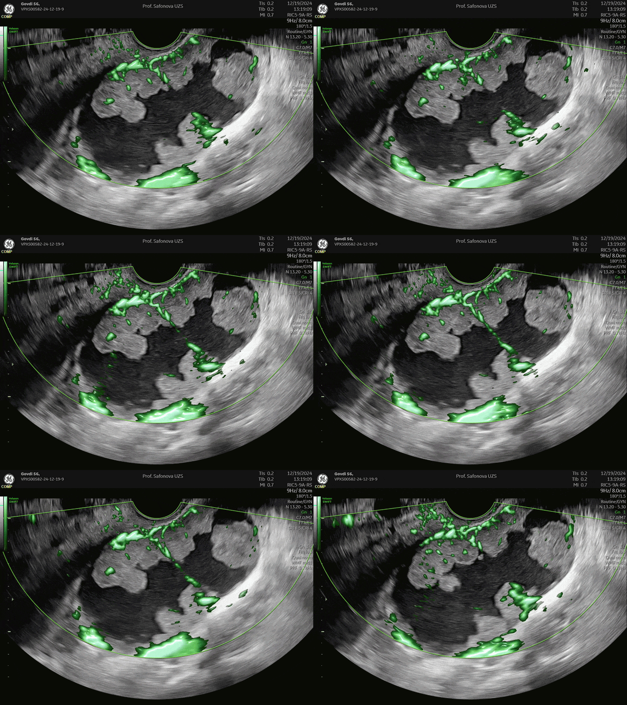
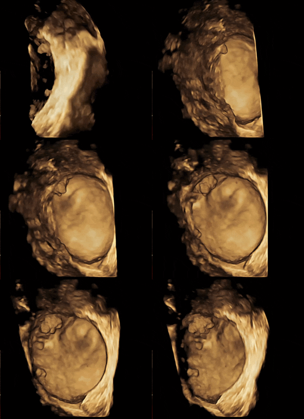
Beyond Static Ultrasound Images
Sono-Endoscopic Correlation with Surgical Findings
Expert Commentary
Concise Reviews of Current Guidelines
Video Clips Instead of Static Images
100+ Clinical Cases with Verified Diagnoses
15+ Hours of Video Content
Annotated Ultrasound Images and Normal Comparisons
Expert Q&A Under Every Lesson
For Professionals at Every Experience Level
Just Getting Started
Build a solid foundation in adnexal ultrasound: examination methodology, anatomy, common benign findings, emergency conditions, and a systematic approach to image interpretation
Have Some Experience
Strengthen the morphological basis of your clinical decision-making. Explore atypical cases, diagnostic pitfalls, and real-world diagnostic errors
Expert
Refine your practice with updated consensus recommendations and contemporary evidence. Explore rare and challenging cases that may surprise even experienced specialists
Sound Familiar?
Why Adnexal Ultrasound Remains Challenging
Similar ultrasound appearances may represent entirely different diagnoses. Conversely, the same pathology can present with different imaging patterns. Diagnostic algorithms may not apply, or may require modification, in special populations such as pregnant women, children, adolescents, and postmenopausal patients
Symptoms and lesion size may not reflect disease severity or prognosis. Many diagnostic errors result from gaps in systematic, up-to-date knowledge and limited experience recognizing uncommon imaging patterns, particularly in rare conditions
Professor Inessa Safonova, MD, PhD Course Director & Instructor
• Over 25 years of experience in ultrasound in obstetrics, fetal medicine, gynecology, and oncogynecology
• ISUOG Ambassador to Eastern Europe
• Senior consultant sonologist at a regional tertiary-level obstetrics and gynecology center, Kharkiv, Ukraine
• Author of 170+ scientific publications and four medical textbooks
• Founder of UZSCHOOL International School of Ultrasound
• Member of the ISUOG Courses Sub-Committee

Three Courses. One Complete Curriculum
44 lessons covering the full spectrum of adnexal ultrasound, from normal findings to oncology
Part 1 - 16 lessons
Normal findings and non-neolpastic lesions
Examination methodology
Anatomy and physiology
Follicles and ovarian morphology
Benign adnexal lesions
Endometriomas
Ectopic pregnancy
Part 2 - 6 lessons
Preoperative risk assessment and US reporting
IOTA Lexicon 2026
O-RADS
GI-RADS, RMI, ROMA, ROCA
Risk stratification
Ultrasound reporting
Pattern recognition
ESGO / ESGE / IOTA / ISUOG consensus 2021
Part 3 - 22 lessons
Benign and malignant adnexal tumors
Benign tumors
Borderline tumors
Malignant tumors
Biomarkers
Ovarian cancer staging
Part 3
Benign and malignant adnexal tumors
Lesson 23
Classifications and Tumor-Specific Ultrasound Features of Adnexal Masses
Classification of ovarian tumors, tumor-specific clinical and ultrasound features, preoperative prediction of histological type, and pattern recognition in ultrasound diagnostics
Lesson 24
Benign Epithelial Serous Ovarian Tumors
Ultrasound presentation of benign serous tumors, including cystadenoma, adenofibroma, and surface papilloma
Lesson 25
Benign Epithelial Mucinous Ovarian Tumors
Recognition and differentiation of benign mucinous ovarian tumors, including mucinous cystadenoma and cystadenofibroma
Lesson 26
Seromucinous Benign Ovarian Lesions
Key diagnostic characteristics of benign seromucinous ovarian lesions, including seromucinous cystadenoma and cystadenofibroma
Lesson 27
Brenner Tumors
Typical presentation of Brenner tumors and the rare occurrence of borderline or malignant forms
Lesson 28
Borderline Epithelial Tumors
Preoperative recognition of borderline ovarian tumors and the distinguishing characteristics of serous, mucinous, and seromucinous subtypes
Lesson 29
Malignant Epithelial Serous Ovarian Tumors
Ultrasound presentation of serous ovarian carcinoma, with attention to low- and high-grade disease and small tumors
Lesson 30
Fallopian Tube Cancer
Ultrasound diagnosis of fallopian tube cancer, illustrated through clinical cases with emphasis on common errors and diagnostic pitfalls
Lesson 31
Malignant Epithelial Mucinous Ovarian Tumors
Recognition of the characteristic morphology and ultrasound presentation of mucinous ovarian carcinomas
Lesson 32
Clear Cell and Endometrioid Carcinoma
Diagnostic characteristics of clear cell and endometrioid ovarian carcinomas, including their association with endometriosis
Lesson 33
Benign Ovarian Stromal Tumors
Ultrasound, clinical, and surgical characteristics of benign sex cord–stromal ovarian tumors
Lesson 34
Malignant Stromal Ovarian Tumors
Ultrasound and surgical characteristics of malignant stromal ovarian tumors, illustrated through clinical scenarios
Lesson 35
Collection of Ovarian Mature Teratomas
Ultrasound appearances of typical, atypical, and difficult-to-visualize dermoid cysts, illustrated through clinical scenarios
Lesson 36
GERM CELL TUMORS. SOME RARE TYPES OF MALIGNANT OVARIAN TUMORS
Key characteristics of benign and malignant germ cell tumors and selected rare types of malignant ovarian tumors
Lesson 37
Metastatic Ovarian Lesions
Clinical presentation and diagnostic characteristics of metastatic lesions involving the ovaries
Lesson 38
Ascites
Ultrasound assessment of ascites, peritoneal carcinomatosis, and other diffuse peritoneal diseases, including clinical significance, disease extent, and prediction of surgical resectability
Lesson 39
Ovarian Cancer: Classification, Staging, Follow-Up
Advanced ultrasound assessment before surgery, classification and staging of ovarian cancer, evaluation of tumor spread, and detection of recurrent disease during follow-up
Lesson 40
Screening or Risk Stratification for Ovarian Cancer
The role and limitations of ovarian cancer screening and the principles of risk stratification
Lesson 41
Adnexal Masses in Postmenopausal Women
Assessment, O-RADS classification, malignancy risk, reporting, and follow-up of simple cysts and complex adnexal masses in postmenopausal women
Lesson 42
Ovarian Lesions in Children and Adolescents
Development, ultrasound diagnosis, and management of pediatric ovarian lesions, including torsion, benign and malignant masses, gonadal dysgenesis, and fertility preservation
Lesson 43
Adnexal Masses in Pregnancy
Diagnosis and management of adnexal masses during pregnancy, including physiological changes, ovarian tumors, endometriomas, torsion, biomarkers, and pregnancy-specific conditions
Lesson 44
Biomarkers of Adnexal Tumors
Selection and interpretation of serological biomarker panels for adnexal tumors in the context of ultrasound findings
How learning works
Full Course Available Immediately
All lessons become available as soon as your enrollment is complete. Study at your own pace and revisit any topic whenever needed
Every Lesson Includes
Video lectures, downloadable presentation slides, and lesson assessments designed to reinforce key concepts and improve retention
Take Notes and Build Your Reference Library
Download presentations, take notes, and keep key classifications, algorithms, and image examples readily available for daily clinical practice
Final Assessment & Certificate*
The course concludes with a final assessment based on ultrasound interpretation and clinical scenarios. Upon successful completion, you will receive a certificate.
Interaction
Expert Q&A Under Every Lesson
Ask questions directly below each lesson. Answers are visible to all participants, building a shared knowledge base
Reference materials
Unlimited Access
Course materials, presentation slides, annotated ultrasound images, and classifications remain available for future reference at any time
Retention
Knowledge Checks
Assess your understanding after each lesson and identify topics that may require further review
Preview Before You Enroll
Inside the Course
Explore preoperative ultrasound findings alongside corresponding surgical and endoscopic images. Learn gynecologic ultrasound pattern recognition through dynamic video clips rather than static screenshots. Review concise case summaries and expert commentary based on real clinical practice
A STORY OF UZSCHOOL
in photos
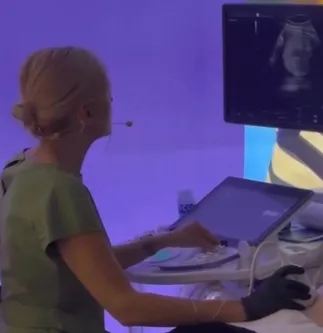
25 years of daily ultrasound experience at a tertiary-level OB/GYN center

With the great P. Jeanty, Kyiv 2018
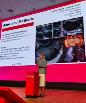
ISUOG World Congress, Budapest 2024
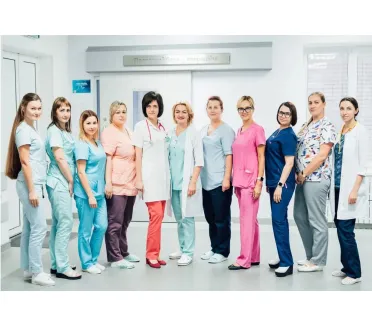
Kharkiv Regional Hospital: Specialized Ultrasound in High-Risk Pregnancy and Women's Health
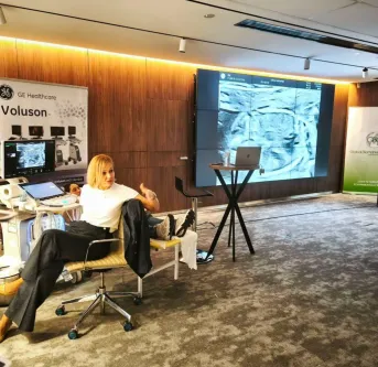
Workshop: live scan and discussion

Ultrasound essentials is a joint collaboration ISUOG and FMF. A session co-chair by invitation of Professor Tom Bourne

UZSCHOOL course in CICE, Clermont-Ferrand, France 2023

UZSCHOOL in Almaty, Kazakhstan 2024

About the school
UZSCHOOL is an international online training center specializing in obstetric and gynecological ultrasound.It serves as a comprehensive guide to OB/GYN for radiologists and a professional pathway into US diagnostics for obstetricians and gynecologists
It all began as a traditional school with in-person training in a conference hall. Since 2019, we’ve transitioned to an online format, which is now recognized by doctors around the world as the most convenient and effective way to learn
5,000+ US specialists from 20+ countries have trained at UZSchool. Participants have joined us from Australia, Azerbaijan, Angola, Armenia, Germany, Georgia, Kazakhstan, Kyrgyzstan, Israel, Spain, Latvia, Lithuania, Moldova, Romania, Poland, Slovakia, Uzbekistan, Ukraine, the Czech Republic, Sweden, Estonia, the United Arab Emirates, Norway and USA
Frequently Asked Questions
Is this course suitable for beginners?
Yes. The curriculum is structured from normal findings to pathology, with examination methodology and key concepts explained step by step
Is this course valuable for experienced practitioners?
Yes. Experienced practitioners often use the course to refine pattern recognition, review current classification systems, and compare ultrasound findings with surgical and endoscopic images
Can I preview a lesson before enrolling?
Yes. You can watch a sample lesson to experience the teaching style, platform features, and course materials before enrolling
Can I study at my own pace?
Yes. All lessons are available immediately after enrollment. However, we recommend following the curriculum sequence, which progresses from physiology and normal findings to oncology
How long will I have access?
Access is unlimited. All lessons, presentations, tests, and supplementary materials remain available in your account for future reference
Choose Your Learning Path
Single Course or the Complete Curriculum
Part 3
120 €
• 22 lessons
• 6+ hours
• 1 year access
• 1 year update
Full Program - Best Value
240 €
• 44 lessons across 3 parts
• 15+ hours
• Lifetime access
• Lifetime update
• Save 60 €